


In this session, we will try to respond to frequently asked questions about PVR examinations. Pulse Volume Recording is a standard and very common test in the physiologic examination of patients with a suspected peripheral vascular disorder.
What is PVR?
PVR, or Pulse Volume Recording, is a non-invasive measurement of the very small changes in the circumference of the limb. These small changes are a function of the blood flow to the limb and can indicate a flow problem in the arteries along the leg.
How is a PVR Test Done?
PVR (Pulse Volume Recording) is a non-invasive test. PVR is also sometimes referred to as Air Plethysmography (APG).
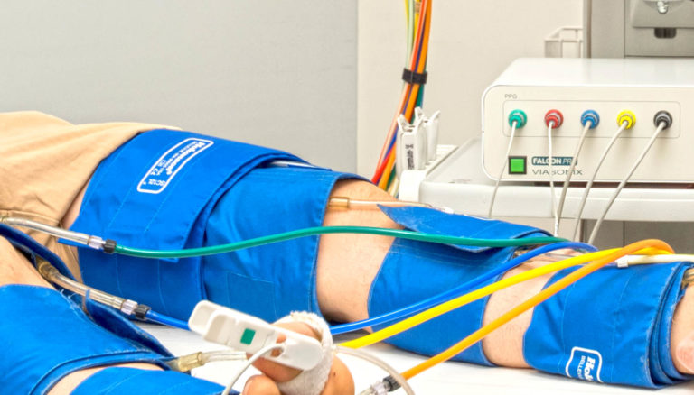
Initially, A pressure cuff is wrapped around the site of interest along the leg. Typically, these sites are the ankle, above or below the knee, or the thigh.
Once the setup is completed, the examiner inflates the pressure cuff to a venous occlusion pressure of about 60 mmHg. From this point, the pressure in the cuff is held constant, and the steady-state waveforms are recorded.
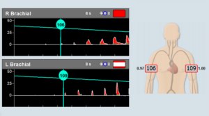
These PVR waveforms indicate very small changes in the air pressure inside the cuff. These minute pressure changes are caused by the subtle limb expansion and contraction as the blood flows through it during systole and diastole.
What is a Normal PVR Waveform?
PVR is a qualitative measurement, which means that the shape of the PVR waveform is reviewed.
The expected results of the PVR waveform for a normal condition are:
- A sharp systolic rise.
- A clear systolic peak.
- A visible dicrotic notch.
- An end-diastolic phase.
What is the Shape of a PVR Waveform?
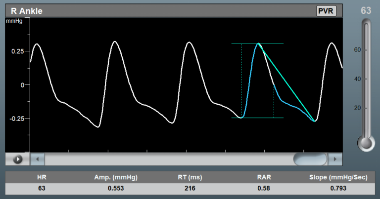
The PVR waveform is an oscillatory signal that changes beat by beat during the cardiac cycle. It normally increases during systole, followed by a decrease in signal amplitude during diastole.
When blood vessels become more rigid as a result of arterial calcification, the peak of the waveform tends to become more rounded with a moderate change in the shape of the peak PVR waveform. In addition, the dicrotic notch, which marks the end of the systole, tends to be less visible and distinguished.
What is Air Plethysmography (APG)?
Air Plethysmography, or APG, means the same as PVR. It is a measure of changes at the site of interest on the limb. APG, or PVR, measures the small limb changes with a pressure cuff filled with air.
As a side note, the other well-known forms of plethysmography are:
- Strain gauge plethysmography.
- Photo-plethysmography.
Is Strain Gauge Plethysmography More Accurate than PVR?
Strain gauge plethysmography (SGP) uses an electrical sensor to measure changes in resistance. The electrical resistance changes reflect changes in strain, force, or pressure.
Typically, a strain gauge is wrapped around the site of interest. In general, strain gauge plethysmography is an old technology rarely used today. PVR (or APG), which is used in machines such as Falcon, is the more common and acceptable clinical method to measure changes in limb circumference or pressure.
Which PVR Parameters are Measured?
The PVR examination is primarily a qualitative diagnosis. However, advanced devices such as the Falcon PVR machine provide additional quantitative measurements. The PVR measurements include:
- The PVR waveform amplitude in units of mmHg,
- The PVR Systolic Rise Time (RT) in units of milliseconds,
- The patient heart rate (HR).
Additional PVR analysis parameters that you should look for are the Relative Amplitude Reduction (RAR) and Down-Slope Curvature (SLOPE or CURV) of the PVR waveform. These parameters are based on Jobst Vascular Laboratory research to characterize vascular disease in diabetic and non-diabetic patients.
Can PVR Measurements be Done Simultaneously?
It is a fundamental vascular requirement that PVR machines will be able to measure multiple PVR sites at the same time. As an example, the Falcon IPU technology (Independent Pneumatic Units) provides the ability to measure up to 10 simultaneous PVR measurements because each pneumatic channel is completely independent.
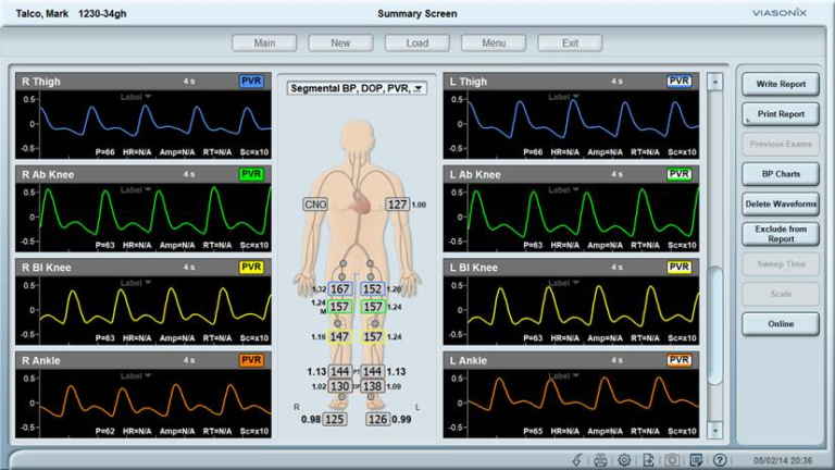
Which Sites are Measured with PVR?
PVR can be measured on any site in the lower or upper limbs. The most common sites for PVR measurements are the ankles, calves, below and above the knees, and the thighs. However, PVR can also be measured on the toes, digits, and above or below the arm elbows.
How Long does it Take to Complete a PVR Test?
In total, the PVR test can take less than 30 seconds per examined site. Pulse Volume Recording is a very short and simple test. It only requires the examiner to inflate a pressure cuff to a low venous occluding pressure, wait for a few more seconds until the PVR waveform stabilizes, and then release the cuff pressure.
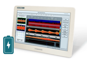
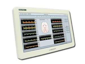
What is the Best PVR Machine?
The Falcon PVR equipment is considered by many as the best PVR machine for numerous reasons.
For example, its IPU technology allows to measure up to 10 PVR sites simultaneously, which saves time. In addition, it displays the waveform in a manner that is ideal for qualitative signal assessment and calculates important quantitative PVR parameters for improved clinical diagnosis. The Falcon also allows performing many other vascular diagnostic examinations that complement the PVR test for a complete vascular assessment. On top of that, the simplicity and user-friendly operation, as well as advanced networking and reporting capabilities, mark the Falcon as the ideal PVR system.
How Much does a PVR Machine Cost?
The general market price of a PVR machine can range from under 15k$ for a basic system without Doppler to 35k$ for a fully loaded PVR device.
The price of a PVR machine depends on its configuration and the number of pressure channels. It also depends on whether the PVR machine includes ultrasound Doppler options or not. As a side note, Doppler is not required for PVR examinations, but it is an integral part of vascular physiologic exams and is normally required by the vascular lab.
What is the Inflation Pressure for PVR Exams?
Depending on the limb and patient condition, the PVR inflation pressure can range between 40mmHg and 80mmHg. Typically, the target PVR inflation pressure is 65 mmHg.
The PVR pressure cuff is inflated to a venous occluding pressure. At this pressure, the cuff inflation is stopped, and after the target pressure stabilizes, the PVR waveform is measured.
What if the Air Pump in the Machine Fails?
A failure of the air pump or any of the other pneumatic components (such as valves) will cause most PVR machines to stop functioning and require service. However, advanced PVR machines have independent pneumatic units (such as IPU Technology). If any component fails, the remaining pneumatic channels continue to function perfectly well. In such a case, there is no immediate rush to perform service on the machine since the patient’s PVR diagnosis can continue without losing a beat.
Is it Important to Color-Code the Pressure Channels?
Color coding of the pressure channels in a PVR machine is essential for the usability of the machine. The examiner can immediately associate the pressure cuff on the limb to a specific pressure channel and to the corresponding on-screen PVR waveform display. It is particularly useful when simultaneous PVR measurements are performed. As an example, the Falcon/PRO PVR machine has 10 color-coded pressure channels, while the Falcon/QUAD and Falcon/ABI+ systems have 4 color-coded pressure channels.