


What is Carotid Endarterectomy?
Carotid Endarterectomy (CEA) is a surgical procedure designed to remove plaque in the Carotid artery by separating it from the carotid arterial wall. Thus, carotid arterial constriction, which may cause a decrease in cerebral perfusion as well as a potential source for cerebral emboli, is removed.
During a CEA procedure, Transcranial Doppler (TCD) monitoring plays a crucial role in ensuring optimal cerebral perfusion and minimizing potential risks.
The Importance of Using TCD in CEA
Transcranial Doppler (TCD) monitoring plays a critical role in Carotid Endarterectomy (CEA) procedures, ensuring optimal outcomes and patient safety. This section highlights the importance of TCD monitoring and its impact on cerebral perfusion and overall procedural success.
Safeguarding Cerebral Perfusion
One of the primary goals of TCD monitoring during CEA is to ensure that cerebral perfusion is not severely impaired throughout the procedure. By continuously assessing blood flow velocities in the intracranial arteries, TCD enables real-time monitoring and early detection of potential issues such as cerebral ischemia or compromised blood flow.
Identifying and Managing Embolic Events
During CEA, the dissection and manipulation of the carotid artery can release emboli that may pose a risk to cerebral circulation. TCD monitoring provides a reliable means of detecting embolic events and quantifying their occurrence. This information enables timely intervention, such as adjusting surgical techniques, implementing protective measures, or initiating appropriate treatments to minimize the impact of emboli on cerebral function.
Guiding Clinical Interventions
TCD monitoring allows medical professionals to make informed decisions and modify the clinical approach in real-time during CEA. For instance, if cerebral ischemia is detected during cross-clamping of the carotid artery, TCD presents real time data to the the surgical team. In such case, the surgical team may decide to intervene and insert a shunt promptly to ensure adequate cerebral perfusion and reduce the risk of complications.
Assessing Collateral Circulation
Evaluation of collateral circulation is vital during CEA to determine if it can provide sufficient protection to the brain in case of temporary reduction in blood flow. TCD monitoring allows the assessment of collateral circulation and provides valuable insights into its capacity and effectiveness. This information helps guide surgical decisions and ensures the safety of the cerebral tissue throughout the procedure.
Enhancing Surgical Precision
TCD monitoring offers real-time data that may assist surgeons in optimizing surgical techniques and achieve precise control during clamping and release, minimizing the risk of ischemic events and promoting favorable patient outcomes.
How to Use TCD for Endarterectomy
This section provides an example of a step-by-step guide on how TCD is utilized to enhance the safety and effectiveness of the procedure. The specific protocol may differ among hospitals and institutions.
Required Equipment

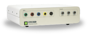
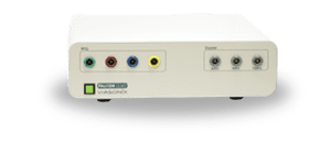
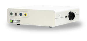
TCD
Machine
Machine

Robotic
Headset
Headset

Manual Headset

2 MHz Doppler Probe
Step 1: Preparing for TCD Monitoring
Before initiating the Carotid Endarterectomy, the TCD monitoring equipment is set up. The system includes a bilateral monitoring headset and preferably a robotic headset, which provides real-time data on cerebral blood flow.
Step 2: Establishing Baseline Measurements
Once the patient is prepared for surgery, baseline measurements of cerebral blood flow parameters are recorded using the TCD system. These parameters include peak and mean blood flow velocities, as well as the pulsatility index (PI) or resistance index (RI).
Step 3: Monitoring During Intraoperative Clamping
As the carotid artery is clamped during the procedure, TCD monitoring allows for continuous assessment of cerebral perfusion. Any signs of cerebral ischemia are closely monitored, indicating the need for intervention to protect the brain. In such cases, the insertion of a shunt may be necessary.
Step 4: Detecting Embolic Events
Throughout the Carotid Endarterectomy, TCD monitoring actively detects and counts suspected embolic events, especially while the clamp is released. With TCD machines, these events are often named HITS (High-Intensity Transient Signals).
Step 5: Real-time Intervention and Assessment
TCD monitoring allows for real-time intervention and adjustments during the procedure. By assessing the collateral circulation and autoregulatory capacity of the circulation, the medical team can modify their approach to minimize risks to the brain.
Using Dolphin TCD for Carotid Endarterectomy
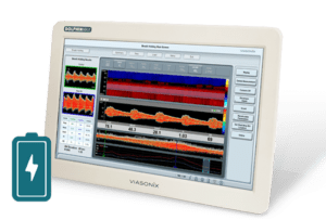
The Dolphin system is an advanced Transcranial Doppler (TCD) monitoring solution that offers numerous benefits when used during CEA. This section highlights a selection of advantages and features of Dolphin in enhancing the use of TCD in the procedure.
- Range of Monitoring Options: The Dolphin system provides a comprehensive range of monitoring options specifically designed for CEA. Alongside the standard bilateral monitoring headset, the Dolphin supports the bilateral Dolphin/XF TCD Robot. This robotic probe scans for intracranial blood flow, assisting physicians in obtaining bilateral cerebral blood flow velocities quickly.
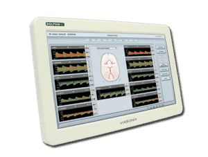
- Real-time Trend Display: During CEA, real-time monitoring of critical parameters is crucial for timely intervention. The Dolphin system offers a clear bilateral trend display of key parameters of interest, such as peak and mean blood flow velocities, as well as the pulsatility index (PI) or the resistance index (RI). This allows immediate assessment and determination of cerebral perfusion during critical stages of the surgery, enabling prompt intervention when necessary.
- High-Resolution HITS Detection: The Dolphin system’s unique high-resolution HITS (High-Intensity Transient Signals) detection capabilities provide exceptional display assisting in identifying embolic events. This feature is particularly relevant during CEA, as embolic showers may occur when the clamp is removed.
- Automatic HITS Event Counting: Suspected embolic events during CEA are automatically counted and displayed on the Dolphin’s screen. This automated counting feature simplifies the monitoring process and allows the surgical team to closely monitor embolic activity and take appropriate measures to minimize their impact on cerebral circulation.
- Power M Mode Display: The Power M Mode display provided by the Dolphin system further enhances the identification of HITS events. This display offers additional visualization and analysis capabilities, supporting the detection and assessment of HITS during the procedure.
Incorporating the Dolphin system for TCD monitoring in Carotid Endarterectomy procedures offers significant benefits. The range of monitoring options, real-time trend display, high-resolution HITS detection, automatic HITS event counting, Power M Mode display, and overall user-friendly design make the Dolphin an invaluable tool in assisting the surgical team during CEA procedures.
Expected Results of CEA with TCD
The primary goal of TCD monitoring is to ensure that cerebral ischemia and associated strokes are prevented. TCD helps identify signs of cerebral ischemia, allowing prompt intervention to protect the brain. Through continuous monitoring, the risk of ischemic strokes associated with CEA can be minimized.
There are various different criteria in the literature to ensure that MCA velocity maintains enough flow during all stages. In some cases, it has been shown that an MCA velocity higher than 10 cm/sec during the critical carotid clamping suggests adequate collateral circulation. On the other hand, intra- and post-operative strokes are associated with a severe (>90%) reduction in velocity with clamping or doubling of the PI (>100%) during the release of clamping.
In addition, post-operative stroke is highly correlated with the amount of TCD detected emboli during CEA.
Selected Literature
Role of transcranial Doppler ultrasonography in stroke, Sanjukta Sarkar et al., Postgrad Med J 2007;83:683–689
Cerebral monitoring during carotid endarterectomy by transcranial Doppler ultrasonography, Woo-Sung Yun, Ann Surg Treat Res 2017;92(2):105-109
Transcranial Doppler ultrasonography in anaesthesia and intensive care, I. K. Moppett and R. P. Mahajan, British Journal of Anaesthesia 93 (5): 710–24 (2004)
Assessment: transcranial Doppler ultrasonography: Report of the Therapeutics and Technology Assessment Subcommittee of the American Academy of Neurology. Sloan MA et al., Therapeutics and Technology Assessment Subcommittee of the American Academy of Neurology. Neurology. 2004 May 11;62(9):1468-81
Transcranial Doppler Monitoring and Causes of Stroke From Carotid Endarterectomy, Merrill P. Spencer, Stroke, Volume 28, Issue 4, April 1997, Pages 685-691
Ultrasound Assessment of the Intracranial Arteries, Nabavi, Ritter, Otis, and Ringelstein, Introduction to Vascular Ultrasonography, By John Pellerito, MD and Joseph F Polak, 2012
Neuro-ultrasonography, Ryan Hakimi, Andrei V. Alexandrov, and Zsolt Garami, Neurol Clin 38 (2020) 215–229
Disclaimer of Information & Content
The content of Viasonix Ltd. website is for information only, not advice or guarantee of outcome. Information is gathered and shared from reputable sources; however, Viasonix Ltd. Management is not responsible for errors or omissions in reporting or explanation. No individuals, including those under our active care, should use the information, resources or tools contained within this self-diagnosis or self-treat any health-related condition. Viasonix Ltd. Management gives no assurance or warranty regarding the accuracy, timeliness or applicability or the content.