Stroke is the result of the death of brain cells due to lack of cerebral perfusion, which in turn causes serious functionality problems that may even lead to death.
The two main types of stroke are:
Transcranial Doppler (TCD) has an established role in the detection and management of stroke patients.
Abnormalities in cerebral blood flow dynamics, as well as abnormalities in blood flow velocities and cerebral vasomotor reactivity, are key in stroke diagnosis and management.
Hemorrhagic stroke, such as during aneurism rupture or subarachnoid hemorrhage (SAH), will result in external compression of the cerebral vasculature, leading to increased cerebral resistance and a significant increase of blood flow velocities due to the decrease in the lumen of cerebral arteries. With an increase in cerebral bleeding, and once the cerebral autoregulatory capacity is exhausted and the arterial lumen further decreases, the blood flow velocities start to decrease sharply. As a result, blood perfusion to the brain tissue is impaired, leading potentially to stroke.
Ischemic stroke can result from various conditions, which cause a reduction in tissue perfusion, such as cerebral emboli. TCD is a preferred modality in identifying and monitoring embolic events and micro-embolic signals, in addition to assessing blood flow abnormalities in the cerebral circulation, and in particular in the arteries that comprise the Circle of Willis.

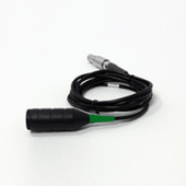
High quality and ultra sensitive Doppler probe

Fast, Stable, and Comfortable Monitoring
The Dolphin is an advanced Transcranial Doppler system that is designed for fast and non-invasive bed-side stroke assessment. The extremely sensitive Doppler probes combined with the high-resolution spectral processing of the Dolphin provide confidence in diagnosing intracranial velocities and waveform dynamics.
Unique trending options allow the immediate display of trends in cerebral blood flow velocities for timely diagnosis of a critical development of cerebral bleeding and patient deterioration.
Velocity threshold markers provide instant and clear indications of vasospasm severity. Various dedicated test protocols, such as breath holding or vasomotor reactivity tests, provide a detailed diagnosis of the cerebral autoregulatory capacity and the vasomotor reactivity.
In addition, the Dolphin has unique abilities in the detection of High-Intensity Transient Signals (HITS), which are suspected embolic events. The extremely high-resolution analysis provides the ability to assess the power and velocity of the suspected emboli, as well as view the suspected emboli traveling along the artery in an exceptional and high temporal resolution m-mode display. Selection of the Power M-Mode display provides an additional display option of the high energetic events in the M-Mode view in addition to the spectral display.
Normal blood flow values of the cerebral vessels comprising the Circle of Willis as well as for the posterior circulation are well documented in the literature. Thus, significantly increased flow velocities suggest the potential for hemorrhagic stroke, while decreased flow that is coupled with abnormal waveform dynamics may lead to ischemic stroke.
The identification of cerebral embolic or micro-embolic signals further indicates the possible type of stroke, leading to a high risk of ischemic stroke.
Role of transcranial Doppler ultrasonography in stroke, Sanjukta Sarkar et al., Postgrad Med J 2007;83:683–689
Trancranial Doppler in stroke, K Rajamani and M Gorman, Biomedicine & Pharmacotherapy, Volume 55, Issue 5, June 2001, Pages 247-257
Assessment: transcranial Doppler ultrasonography: Report of the Therapeutics and Technology Assessment Subcommittee of the American Academy of Neurology. Sloan MA et al., Therapeutics and Technology Assessment Subcommittee of the American Academy of Neurology. Neurology. 2004 May 11;62(9):1468-81
Transcranial Doppler: Techniques and advanced applications: Part 2, Sharma AK, Bathala L, Batra A, Mehndiratta MM, Sharma VK, Ann Indian Acad Neurol. 2016 Jan-Mar;19(1):102-7
Ultrasound Assessment of the Intracranial Arteries, Nabavi, Ritter, Otis, and Ringelstein, Introduction to Vascular Ultrasonography, By John Pellerito, MD and Joseph F Polak, 2012
Neuro-ultrasonography, Ryan Hakimi, Andrei V. Alexandrov, and Zsolt Garami, Neurol Clin 38 (2020) 215–229
Transcranial Doppler, Peter J. Kirkpatrick and Kwan-Hon Chan, Chapter 13, in Head Injury. Edited by Peter Reilly and Ross Bullock. Published in 1997 by Chapman & Hall, London
Disclaimer of Information & Content
The content of Viasonix Ltd. website is for information only, not advice or guarantee of outcome. Information is gathered and shared from reputable sources; however, Viasonix Ltd. Management is not responsible for errors or omissions in reporting or explanation. No individuals, including those under our active care, should use the information, resources or tools contained within this self-diagnosis or self-treat any health-related condition. Viasonix Ltd. Management gives no assurance or warranty regarding the accuracy, timeliness or applicability or the content.Contact us via email: [email protected]
Or using the form below.
Our team will respond back quickly!