


What is Spectral Doppler of Physiologic Machines?
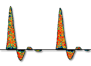
Spectral Doppler is the outcome of sophisticated algorithms and analysis of the Doppler signals based on FFT analysis (Fast Fourier Transform). This algorithm results in a color-coded display of the blood flow velocities within the vascular lumen as a function of time. The scatterers within the blood vessel, typically the red blood cells (RBC), are traveling at varying velocities. The color code maps the different concentrations of the RBCs that are moving at each specific velocity range.
What are the Doppler Envelope, the Peak, and Mean Blood Flow Velocities?
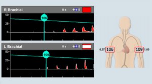
The Doppler envelope is the outline of the peak blood flow velocity as a function of time.
Visually, it is similar to drawing a line along the external boundaries of the Doppler spectrum. Within each cardiac cycle, the highest envelope value represents the peak blood flow velocity. The average of the envelope over the cardiac cycle represents the mean blood flow velocity.
What is the Clinical Importance of Spectral Doppler Analysis?
How to use Spectral Doppler Analysis with ABI (Ankle Brachial Index)?
First, the examiner wraps a cuff around the segment of the arm or limb to be tested and uses the Doppler probe (most commonly 8 MHz Doppler probe) to locate an artery and measure it. The examiner then inflates the cuff and attempts to reach occlusion. Finally, the systolic blood pressure is defined as the point in time that the Doppler signal returns as the occluding pressure cuff is slowly deflated.
How to use Spectral Doppler Analysis for Arterial Diagnosis?
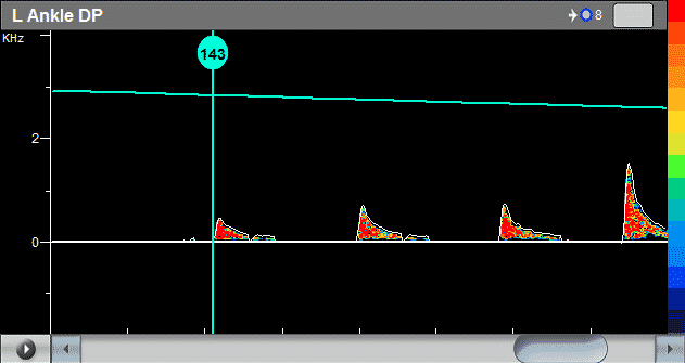
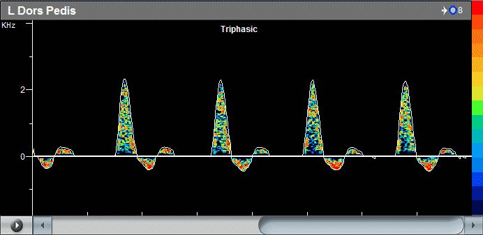
During the routine vascular examination, the examiner uses Doppler measurements to determine the vasculature state. Doppler measurements, in this case, are typically done in the lower extremities in the Anterior Tibial artery, Dorsalis Pedis artery, Popliteal artery, and Femoral artery. The primary objective is to determine whether the Doppler waveform is monophasic, biphasic, or triphasic (and even multi-phasic waveform).
While tri-phasic waveforms suggest that the arteries are healthy and elastic, the loss of Doppler phases may indicate calcification and hardening of the arteries that may lead to other pathological conditions.
Other uses of Doppler measurements include hemodynamic assessment of the flow, including flow disturbance, systolic window assessment, and wave reflections. Doppler can also be used in various specific vascular specialty tests, including impotence evaluation, Thoracic Outlet Syndrom diagnosis, Palmar Arch testing, and more.
Why is Spectral Doppler clinically superior to just displaying the envelope?
The exact timing and location of the initial appearance of the Doppler signal after occlusion and deflation of the pressure cuff are critical in the correct measurement of the systolic blood pressure. Any error in the determination of the ankle or brachial pressures may significantly affect the calculation of the ABI index, potentially leading to incorrect clinical diagnosis. With the spectral Doppler display, it is completely clear to the examiner when the Doppler signal returns, even when the signal is very low in amplitude. In contrast, when only the envelope is displayed, and the amplitude is, low the general Doppler noise and the envelope get mixed and make it difficult to determine the exact systolic pressure.
Similarly, during routine Doppler measurements for assessing the phasic nature of the flow, it is critical to determine the number of Doppler waveform phases correctly. Typically, the second and, more importantly, the third phase are significantly smaller in amplitude than the first phase. With a color spectrum, the different phases are easily seen, and the correct diagnosis is straightforward. However, when only the envelope is displayed, any noise interference can corrupt the correct count of the Doppler phases.
Finally, hemodynamic assessment of the Doppler blood flow velocity is possible only with spectral Doppler. Phenomena such as systolic bruit, turbulence, or “systolic window” are clearly visualized in the color Doppler display. The envelope outlining the velocity provides no information about the hemodynamics taking place inside the vascular lumen.
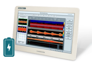
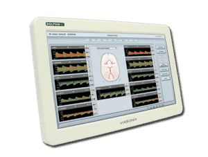
What is the best physiological machine with the best spectral Doppler display?
The Falcon is considered to be the best physiological machine, and it has a crystal clear Spectral Doppler display. The Falcon is the only advanced physiological testing machine in the USA with real and complete Doppler spectrum analysis and display. While other devices display just the Doppler envelope without the tools to determine the clinical validation of the measurements, the Falcon stands out as the leading Doppler system.
The Spectral Doppler display of the Falcon device allows clinicians to feel completely comfortable with their measurements and vascular diagnosis. Furthermore, detailed analysis and diagnosis that are not possible with other vascular machines that do not have full-color spectral display are made possible with the Falcon!
Falcon Doppler probes are made of the highest quality, and together with the advanced spectral analysis, provide the optimal clinical, physiologic machine for vascular diagnosis.
For more information, please leave us a message.