


What is Extracranial Exam?
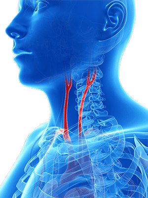
Extracranial Doppler examinations are measurements of blood flow velocities in the extracranial vessels, and particularly the Common, External, and Internal Carotid arteries, as well as the Subclavian artery. These measurements are key in the diagnosis of cerebral pathology and circulation.
How to Perform Extracranial Tests
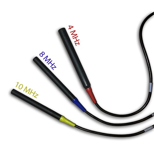
The extracranial vessels are easily accessible with standard Continuous Wave (CW) Doppler probes and allow quick identification of abnormal blood flow patterns. The selection of the Doppler probe frequency depends on the vessel size and its’ distance from the skin. The 4 MHz and 8 MHz frequencies are the most common selections for extracranial measurements.
Extracranial measurements serve various clinical purposes, such as identifying carotid stenosis or similar lesion, determining increased distal cerebral resistance, Subclavian Steal Syndrome, and Stroke assessment. The measured blood flow velocities increase significantly in the area of stenosis, until the stenosis decreases the effective arterial diameter to a critical level.
Another common parameter is the Lindegaard Ratio (LR). The LR is defined as the ratio between the mean Middle Cerebral artery (MCA) velocity and the mean Internal Carotid Artery (ICA) velocity. It is an essential diagnostic parameter when determining if high cerebral velocities result from a cerebral hemorrhage, arterio-venous malformations, or other factors.
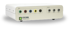
Doppler Probes
Essential for Carotid Measurements
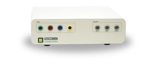
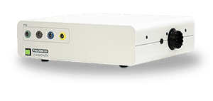
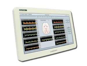
Using the Falcon for Extracranial Measurements
The Falcon supports a variety of CW Doppler probes, including 4 MHz, 8 MHz, and 10 MHz probes.
These frequencies allow optimal access and measurements of larger/smaller and deeper/superficial blood vessels. The higher frequencies are intended for smaller and shallower vessels, and vice versa.
The Falcon protocols allow the configuration of any blood vessel of interest, and this is supported with a schematic picture of the carotid circulation for improved documentation.
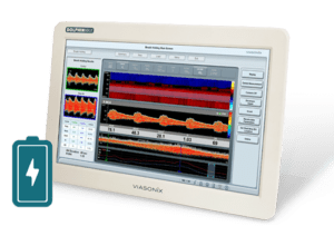
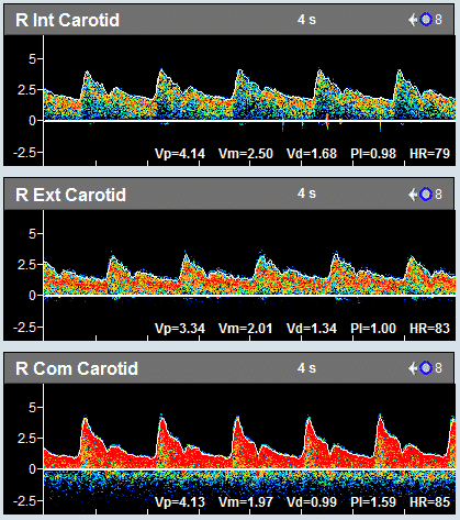
The Doppler measurements include complete spectral analysis, which allows optimal diagnosis and visualization, for example, of systolic bruits. A complete set of Doppler parameters is automatically calculated, including:
- Peak velocity,
- Mean velocity,
- Diastolic velocity,
- Pulsatility index (PI),
- Resistance index (RI),
- Systolic-Diastolic ration (S/D), systolic rise time, and
- heart rate.
- The spectral color palette, display, and noise rejection can be configured and optimized for each user.
Likewise, the peak systolic envelope can be controlled, Y-Axis units (cm/sec or KHz), as well as a range of other Doppler controls, including Sweep Time, Gain, High-Pass Filter, Scale, and Volume.
Expected Results
The criteria for the assessment of the extracranial blood flow measurements depends on the pathology. Focal stenosis will cause a significant increase in mean and peak blood flow velocities, up to a critical level, after which flow will decrease. An intracranial hemorrhage and increased MCA velocity will likely result in higher Lindegaard ratio values. Other criteria for the assessment of pathology are further defined in the accepted international guidelines.
Selected Literature
Cerebrovascular Ultrasound in Stroke Prevention and Treatment, Edited by Andrei V. Alexandrov, Blackwell Publishing 2004
Ultrasound Assessment of the Intracranial Arteries, Darius G. Nabavi et al., in “Introduction to Vascular Ultrasonography”, Ed. Pellerito and Polak, Elsevier Health Sciences, 2012, Ch 12, pp 202-228
Transcranial Doppler, Peter J. Kirkpatrick and Kwan-Hon Chan, Head Injury. Edited by Peter Reilly and Ross Bullock. Published in 1997 by Chapman & Hall, Ch 13, pp 243-259
Disclaimer of Information & Content
The content of Viasonix Ltd. website is for information only, not advice or guarantee of outcome. Information is gathered and shared from reputable sources; however, Viasonix Ltd. Management is not responsible for errors or omissions in reporting or explanation. No individuals, including those under our active care, should use the information, resources or tools contained within this self-diagnosis or self-treat any health-related condition. Viasonix Ltd. Management gives no assurance or warranty regarding the accuracy, timeliness or applicability or the content.