


What is Pulse Wave Velocity?
Pulse Wave Velocity (PWV) is a simple and non-invasive measurement, which can be measured at various locations along with the arterial circulation to assess arterial stiffness.
Pulse wave velocity (PWV) is widely recognized as a simple and reliable clinical measure of arterial stiffness/elasticity, which is correlated with vascular disease. The contractions of the heart, which drive the arterial blood, also generate arterial blood pressure pulse waves which propagate through the arterial walls. PWV is defined as the velocity at which these arterial blood pressure pulses propagate.
How to Measure Pulse Wave Velocity
While there is no one set standard of how or where to measure PWV, some of the more common PWV measurements include the baPWV, which reflects the pulse wave propagation between the brachial and ankle, cfPWV reflecting propagation between the carotid and femoral, and faPWV also know as leg PWV reflecting the PWV along the leg between the femoral and ankle.
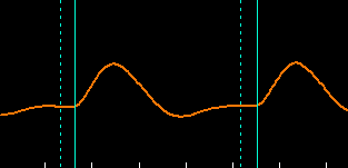
The measurement of PWV is usually a straightforward calculation based on the definition of velocity, i.e., distance divided by time. A physiologic signal, typically the initial systolic upstroke, serves as a time marker. When the arterial pulse wave propagates along with the arterial circulation, a short time delay is generated between the proximal and the distal measurement sites. The distance between the 2 measurement sites can be either measured directly or estimated based on some parameters such as patient height. When the PWV is not measured along a straight arterial line, such as the cfPWV, various assumptions need to be made.

Inflatable Cuffs
High quality, available in a variety of sizes
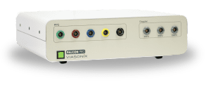
Using the Falcon for PWV measurements
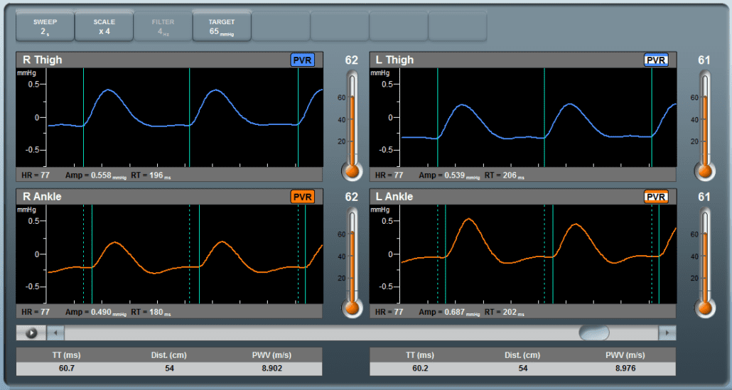
The Falcon has a dedicated PWV protocol, allowing a simple and non-invasive diagnosis of various PWV parameters such as the baPWV or faPWV. The measurement is performed with the aid of blood pressure cuffs. These cuff are wrapped around the target measurement sites, such as:
- The Ankle and Thigh for faPWV, or
- Brachial and ankle for baPWV.
The patient is in the supine position during the measurement, and the pressure cuffs are inflated to a venous occlusion pressure, typically around 65 mmHg. The pressure is kept steady while the corresponding PVR waveforms are displayed as a function of time. The examiner can determine the target inflation pressure.
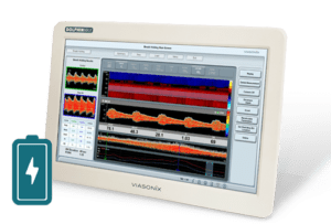
Typically, the examination lasts a few minutes, during which multiple measurements for each cardiac cycle are performed, and the time delay between the 2 measurement cuffs is automatically determined. The distance between the pressure cuffs is entered manually or calculated automatically based on known relationships. Once the examiner stops the measurement process, PWV is calculated automatically and displayed on the screen as an average of the measurements over the approved cardiac cycles.
The Falcon allows to perform the PWV test bilaterally, i.e., simultaneous measurements for the right and left sides. Automatic time markers clearly display the time gap between the proximal and distal systolic rise time to verify the measurement process.
Expected Results
PWV is a quantitative parameter reflecting arterial stiffness or elasticity. Higher PWV values reflect greater than normal arterial stiffness.
This parameter is highly dependent on the method of measurement, as well as on the measurement sites. Therefore, typical values for baPWV differ from faPWV and cfPWV.
In addition, PWV is highly dependent on patient age. Older patients are expected to have more stiff arteries and, as a result, higher PWV values under normal conditions. Diabetic patients are also expected to have a significant increase in PWV values.
Based on literature reports, normal PWV values can range between around 6 m/s to 14 m/s, depending on the method of measurement and the patient’s age.
Selected Literature
Familial Tendency for Hypertension is Associated with Increased Vascular Stiffness, Wexler et al., Journal of Hypertension 2020, 38.
Peripheral vascular disease assessment in the lower limb: a review of current and emerging non‑invasive diagnostic methods; Shabani Varaki et al, BioMed Eng OnLine (2018) 17:61
Determinants of pulse wave velocity in healthy people and in the presence of cardiovascular risk factors: ‘establishing normal and reference values’, Boutouyrie et al., European Heart Journal (2010) 31, 2338–2350
Brachial–ankle pulse wave velocity: an index of central arterial stiffness?, Sugawara et al., Journal of Human Hypertension;19, 401–406, February 2005
Arterial path length estimation on brachial-ankle pulse wave velocity: validity of height-based formulas, Sugawara et al., Journal of Hypertension 2014, 32:881–889
Arterial stiffness predicts cardiovascular outcome in a low-to moderate cardiovascular risk population: the EDIVA (Estudo de DIstensibilidade VAscular) project, Joao Maldonado et al., Journal of Hypertension 2011, 29:669–675
Effects of aging on leg pulse wave velocity response to single-leg cycling, Sugawara et al., Artery Research (2010) 4, 94e97
Association between estimated pulse wave velocity and the risk of stroke in middle-aged men, Sae Young Jae et al., International Journal of Stroke, 0(0) 1–5, 2020.
Disclaimer of Information & Content
The content of Viasonix Ltd. website is for information only, not advice or guarantee of outcome. Information is gathered and shared from reputable sources; however, Viasonix Ltd. Management is not responsible for errors or omissions in reporting or explanation. No individuals, including those under our active care, should use the information, resources or tools contained within this self-diagnosis or self-treat any health-related condition. Viasonix Ltd. Management gives no assurance or warranty regarding the accuracy, timeliness or applicability or the content.