What are Segmental Blood Pressures?
Lower extremity Segmental Blood Pressures (SBP) diagnosis refers to the measurement of the systolic blood pressure at various sites along each leg.
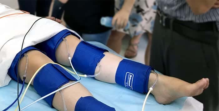
Segmental blood pressures is a physiologic test, which is performed using a physiologic machine in order to help in localizing arterial obstruction to flow along the limb, as well as the physiological severity of the obstruction. This is in contrast to ultrasound imaging of the lower limb, which is focused on detecting and quantifying the anatomical severity of the arterial obstruction to flow.
Physiologic and anatomical two types of tests complement each other. They are frequently performed one after the other to diagnose both the physiological and the anatomical severity of the pathology for optimal clinical diagnosis.
Segmental Blood Pressures typically include the following lower extremity sites:
- Thigh
- Above knee
- Below knee
- Ankle
- Sometimes calf, metatarsals, and toes are also included.
How to Perform Segmental Blood Pressures Test
Measuring segmental blood pressures is a crucial part of the diagnostic process for evaluating lower extremity vascular health. The procedure requires the use of a specialized segmental pressures machine, which is designed to provide fast and accurate measurements.
Here is a detailed guide on how to perform the segmental blood pressure test:
- Preparation:
- Ensure that the patient is in a comfortable position, preferably lying down or in a supine position, to promote accurate measurements and minimize movement artifacts.
- Expose the lower extremities, including both legs, from the thigh down to the toes, for cuff placement and Doppler probes or PPG sensor application. Note that Doppler is superior and is widely considered the gold standard method.
- Equipment Setup:
- Select the appropriate cuff sizes for each target site along both legs. The cuffs should fit snugly but not too tight to avoid interference with blood flow.
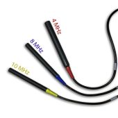
- Attach Doppler probes with frequencies (e.g., 4MHz, 8MHz, 10MHz) suitable for the target vessels such as Posterior Tibial or Dorsalis Pedis arteries.
- Position PPG sensors on the toes for additional measurements.
- Cuff Placement:
- Begin the measurement process at the ankle level by placing cuffs around the ankles of both legs. Ensure that the cuffs are correctly positioned and secure.
- Gradually move upward along the leg, placing cuffs at the above knee, below knee, and thigh sites, if necessary.
- Inflation and Measurement:
- Inflation: Inflate each cuff while observing the downstream Doppler or PPG waveform. The inflation pressure should exceed the arterial systolic pressure.
- Deflation: Gradually bleed the cuff pressure until the distal Doppler or PPG waveforms reappear. The pressure at the initial re-appearance is considered the systolic pressure.
- Repeat and Record:
- Repeat the inflation and deflation process for each cuff on both legs to obtain a comprehensive set of measurements.
- Record the systolic pressure values for each site, including ankle, above knee, below knee, and thigh, for further analysis.
- Interpretation:
- Analyze the recorded pressures to determine pressure differences between adjacent sites and between similar sites on both legs.
- A relatively large pressure difference (pressure drop) may suggest the presence of arterial obstruction at specific locations.
- Additional Considerations:
- Ensure that the Doppler and PPG sensors are positioned correctly and providing clear waveforms during the measurement process.
- Pay attention to any potential occlusion effects on the complete limb and take measures to minimize them.
- Utilize the features and options of the segmental pressures machine, such as automatic cuff inflation and simultaneous measurements, for a faster and efficient test process.
This step-by-step guide for performing a Segmental Blood Pressures test is for informational purposes only. Healthcare professionals should rely on their expertise, clinical judgment, and institutional protocols for accurate administration and interpretation. Additional clinical information, patient history, physical examination, and other diagnostic tests may be necessary for comprehensive evaluation.
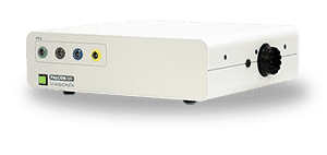
Using the Falcon for SBP Test
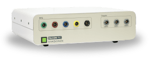
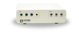
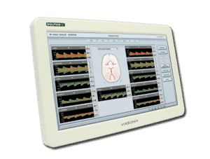
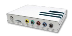
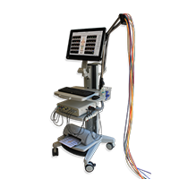
The Falcon segmental pressures machine offers numerous advantages and benefits when conducting the segmental blood pressure test. Specifically, the Falcon/Pro is often considered the ideal model to use, as it has 10 separate color-coded pressure channels designed to simplify the examination process.
Designed to be practical and efficient, the Falcon can potentially enhance the diagnostic process for healthcare professionals and benefit patients. Here are some key potential benefits of using the Falcon for the SBP test:
- Rapid and Efficient Testing:
The Falcon/Pro model is equipped with 10 separate color-coded pressure channels, enabling simultaneous measurements at multiple sites along both legs. As a result, the test can be completed quickly, saving valuable time for both healthcare providers and patients. - Wide Range of Sensors:
With a wide range of Doppler probes of various frequencies (4MHz, 8MHz, 10MHz) and color-coded PPG sensors (Disk, toe clips, finger clips), the Falcon ensures optimal probe and sensor selection. - User-Friendly Interface:
The Falcon’s user-friendly interface simplifies the testing process and can minimize the learning curve for healthcare practitioners.
Customizable options, such as target inflation pressure, deflation rate, sweep time display, gain, and filter, allow for tailored testing according to patient needs. - Comprehensive Diagnostics:
Sequentially inflating and deflating each ipsilateral pressure cuff along the leg allows for localization of arterial obstructions along the limb. Because of that, the Falcon complements ultrasound imaging by focusing on physiological severity rather than just anatomical severity of arterial obstructions (1, 2, 3, 4). - Enhanced Patient Comfort:
High-quality inflatable cuffs are available in various sizes, to help ensure a comfortable and secure fit during the test. The test can be conducted while the patient is in a supine position, promoting relaxation and reducing potential movement artifacts. - Advanced Features:
The Falcon offers numerous features, including automatic cuff inflation as soon as a signal is identified, simultaneous measurements, and display of contralateral results. Most importantly, an automatic cursor placed at the proposed systolic pressure location allows for manual adjustments and improved measurement accuracy as the examiner finds fit. - Data Analysis and Documentation:
The Falcon automatically displays the pressure index for each site, facilitating data analysis and comparison between right and left brachial pressures. Additionally, comprehensive results and analysis can help to simplify the documentation and reporting for further clinical evaluation. - Versatile and Suitable for Various Clinical Settings:
The Falcon/Pro model is designed for use in large medical institutions as well as smaller clinics. Its versatility, ease of use, and rapid results make it valuable for routine day-to-day work and other clinical applications.
Expected Results of Segmental Blood Pressures
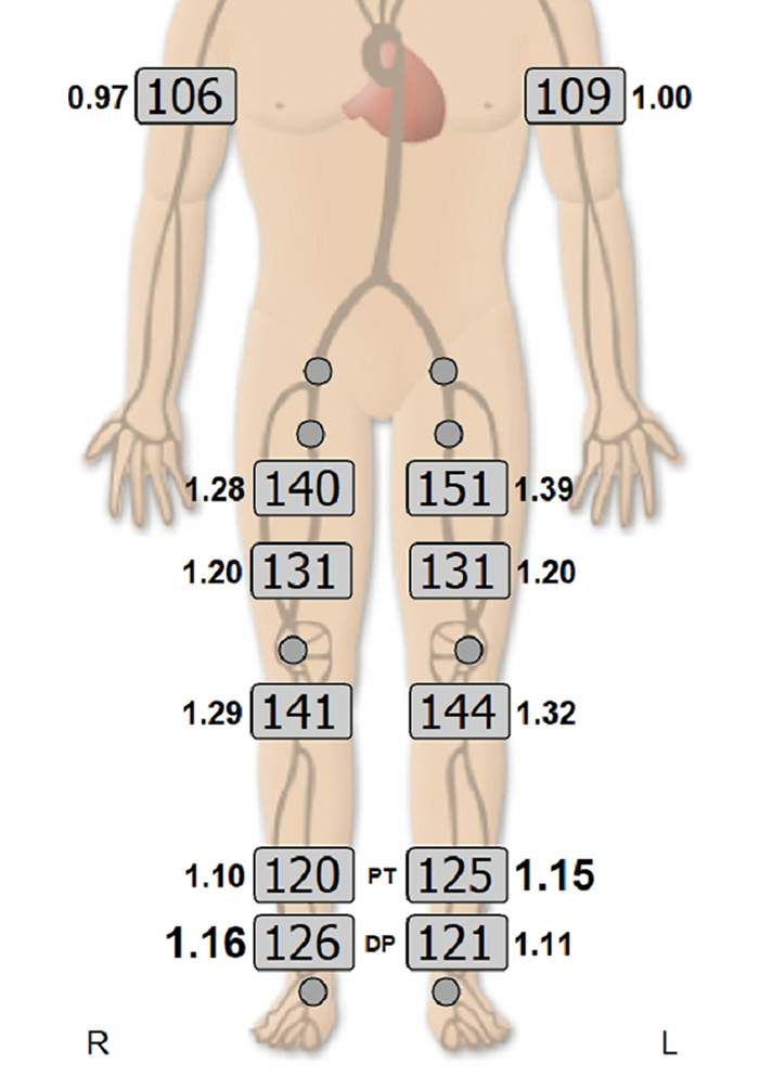
When measuring segmental blood pressures, the focus is on the pressure difference between each 2 consecutive adjacent sites, as well as the pressure difference between similar sites on the right and left legs (2).
A relatively large pressure difference (or pressure drop) suggests the location of the pathology (2). For example, a large pressure drop between the thigh and the adjacent above knee site suggests an arterial obstruction to flow, which is located between the thigh and the knee.
The magnitude of the pressure drop threshold that marks a pathological condition may vary from one institute to another. Still, a common thumb rule is a pressure difference which is typically larger than 20 mmHg (2).
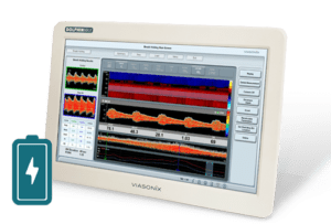
An example of a SBP measurement taken from Viasonix Falcon/PRO physiologic machine using 8 MHz CW Doppler probe.
Selected Literature
- Overview of Peripheral Arterial Disease of the Lower Extremity, Ali F. AbuRahma and John E. Campbell, Noninvasive Vascular Diagnosis, A.F. AbuRahma (ed.), Springer International Publishing AG 2017, Ch 21, pp 291-318
- Peripheral vascular disease assessment in the lower limb: a review of current and emerging non‑invasive diagnostic methods, Shabani Varaki et al, BioMed Eng OnLine (2018) 17:61
- 2016 AHA/ACC Guideline on the Management of Patients With Lower Extremity Peripheral Artery Disease; A Report of the American College of Cardiology/American Heart Association Task Force on Clinical Practice Guidelines; Journal of the American College of Cardiology, Vol. 69, No. 11, 2017
- Lower Extremity Arterial Physiologic Evaluations, Vascular Technology Professional Performance Guidelines, the Society for Vascular Ultrasound, 2019
Disclaimer of Information & Content
The content of Viasonix Ltd. website is for information only, not advice or guarantee of outcome. Information is gathered and shared from reputable sources; however, Viasonix Ltd. Management is not responsible for errors or omissions in reporting or explanation. No individuals, including those under our active care, should use the information, resources or tools contained within this self-diagnosis or self-treat any health-related condition. Viasonix Ltd. Management gives no assurance or warranty regarding the accuracy, timeliness or applicability or the content.
