


What is Thoracic Outlet Syndrome?
The Thoracic Outlet Syndrome test (TOS) is an examination performed primarily to discriminate between the causes of patient symptoms due to compression at the thoracic outlet. The compression can affect the blood vessels or nerves, and the TOS test is conducted to determine whether the patient symptoms originate from vascular or neurogenic causes.
Indications for TOS Test
The Thoracic Outlet Syndrome (TOS) test is a crucial tool for diagnosing patients with potential compression at the thoracic outlet. It is recommended when patients exhibit:
Upper Extremity Symptoms: TOS testing is relevant for those with pain, numbness, tingling, weakness, or skin color changes in the arms, hands, or fingers.
Neurological Symptoms: Patients showing abnormal sensations, muscle weakness, or reduced reflexes in upper limbs may need a TOS test due to possible nerve compression.
Vascular Symptoms: A TOS test benefits those with coldness, discoloration, or altered pulse in the upper extremities, indicating vascular compression and reduced blood flow.
Aggravating Factors: Symptoms worsening with arm raising, head tilting, or specific activities suggest thoracic outlet compression.
Occupational or Lifestyle Factors: Those with jobs involving repetitive overhead motions or prolonged arm positioning could require TOS testing if symptomatic.
History of Trauma: Patients with neck, shoulder, or upper chest trauma history are at higher risk and could benefit from a TOS test.
Failed Conservative Management: If upper extremity symptoms persist despite conservative treatments, a TOS test can evaluate potential thoracic outlet compression.
Differential Diagnosis: Patients with TOS-like symptoms should undergo testing to distinguish from other causes like cervical spine disorders or nerve entrapment.
Note: Clinical judgment of healthcare professionals guides the decision for TOS testing. Comprehensive evaluation, including medical history, physical exam, and other tests, is essential to accurately identify candidates for the TOS test.

How to Perform Thoracic Outlet Syndrome Test
Performing a TOS test requires proper technique and adherence to established protocols.
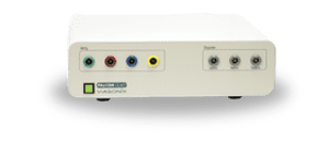
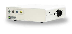
Required Equipment
Vascular
Machine
Machine
PPG
Sensors

Doppler
Probes
Inflatable Cuffs
Here is a step-by-step guide on how to perform a TOS test:
Step 1: Introduction and Setup
- Prepare the necessary equipment, including a diagnostic system capable of measuring various parameters like Doppler, PVR (Pulse Volume Recording), and PPG (Photoplethysmography).
- Ensure the patient’s comfort and proper positioning for the examination.
- Select a TOS protocol in your vascular diagnostics equipment.
- Position PPG sensors on both the right and left digits.
- Alternatively, you can also use a Doppler probe to measure one of the hands, or PVR cuffs to measure both hands. The rest of this guide assumes that PPG sensors are used.
Step 2: Initial Resting Measurements
- Use the PPG sensors to measure normal resting waveforms in the digits of the patient. This establishes a baseline for comparison.
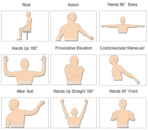
- Positional Maneuvers: Guide the patient through a series of positional maneuvers to assess perfusion and symptoms. These maneuvers help identify if specific positions result in abnormal decreases in perfusion, indicating a potential vascular disorder. The sequence of maneuvers can include:
- Hands-up Test: The patient raises their hands.
- Adson or Scalene Maneuver: The patient tilts their head and turns it towards the examined shoulder.
- Costoclavicular Maneuver: The patient pulls their shoulders back while pushing their chest forward.
- Provocative Elevation Test: The patient elevates their arms.
- And more maneuvers: As necessary, based on clinical judgment.
- Symptomatic Position Identification: The focus of the examination is to identify a position that significantly reduces perfusion and reproduces the patient’s symptoms. It is advised to document the symptomatic position using a camera or other suitable means. This position is crucial for diagnosis.
Step 3: Quantitative and Qualitative Analysis
- Evaluate the measured waveforms, both from the right and left sides, for each positional maneuver.
- Look for abnormal decreases in amplitude or signal flattening that correspond to the symptomatic position.
- The diagnosis primarily involves visual assessment, but quantitative measurements such as PPG, PVR or Doppler waveform amplitudes can support the findings.
Step 4: Diagnosis and Documentation
- Based on the visual and quantitative analysis, determine whether the symptoms originate from a vascular disorder.
- Document the findings, including the symptomatic position, waveform characteristics, and any abnormalities observed.
- Interpret the results in the context of the patient’s clinical presentation.
- Prepare a report summarizing the examination, findings, and conclusions.
This step-by-step guide for performing a TOS test is for informational purposes only. Healthcare professionals should rely on their expertise, clinical judgment, and institutional protocols for accurate administration and interpretation. Additional clinical information, patient history, physical examination, and other diagnostic tests may be necessary for comprehensive evaluation.
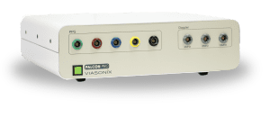
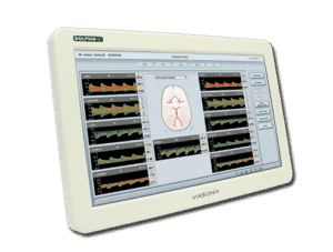
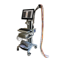
Using the Falcon for TOS Assessment
The Falcon system revolutionizes Thoracic Outlet Syndrome (TOS) diagnosis, offering multiple benefits for healthcare professionals and patients.
Tailored with a dedicated TOS protocol, Falcon optimizes efficiency in TOS testing, presenting the following key advantages:
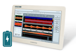
Precise Visual Protocol: Falcon’s TOS protocol includes visual guides, aiding examiners and patients through positional maneuvers with schematic images.
Customize Maneuvers: While including a visual step-by-step protocol with the essential maneuvers, Falcon also allows customization with additional maneuvers.
Symptomatic Position Documentation: Falcon’s built-in camera captures the symptomatic position, integrating it into the protocol and reports for precise documentation.
Comprehensive Testing: Falcon supports various TOS tests, including Doppler and PVR measurements alongside standard PPG sensors.
Efficient Multi-Site Measurement: The Falcon/Pro enables simultaneous measurement of up to 5 PPG sensors and up to 10 PVR waveforms per finger.
Quantitative Parameters: Falcon provides quantitative measurements like PPG and PVR waveform amplitudes and Doppler parameters to aid with the interpretation and documentation.
Streamlined Reporting: Falcon’s documentation of positions, waveforms, and abnormalities simplifies examination reports, facilitating communication among healthcare professionals.
Optimized Workflow: Falcon’s user-friendly interface and automated data collection streamline testing, benefiting professionals and patients by reducing examination time.
Expected Results
The diagnosis of the TOS test is primarily qualitative in nature. The focus is on viewing the measured waveforms on both the right and left sides for each positional maneuver and determining whether a specific position resulted in an abnormal decrease in amplitude or resulted in signal flattening. Such a case is indicative of a potential decrease in perfusion, suggesting a vascular source of TOS.
Selected Literature
Reporting standards of the Society for Vascular Surgery for thoracic outlet syndrome, Karl A. Illig et al., J Vasc Surg 2016;64:e23-e35.
Diagnosis and Treatment of Thoracic Outlet Syndrome, Editors Julie Ann Freischlag and Natalia O. Glebova, 2018 MDPI, Basel, Switzerland
Disclaimer of Information & Content
The content of Viasonix Ltd. website is for information only, not advice or guarantee of outcome. Information is gathered and shared from reputable sources; however, Viasonix Ltd. Management is not responsible for errors or omissions in reporting or explanation. No individuals, including those under our active care, should use the information, resources or tools contained within this self-diagnosis or self-treat any health-related condition. Viasonix Ltd. Management gives no assurance or warranty regarding the accuracy, timeliness or applicability or the content.