


What is Intracranial Stenosis?
Intracranial Stenosis is a narrowing of a cerebral artery, which can develop primarily due to intracranial atherosclerosis or acute thrombus. It is a significant risk factor for Transient Ischemic Attack (TIA) and ultimately a Stroke.
How to use TCD for Intracranial Stenosis Test
Transcranial Doppler is widely used to detect and determine the severity of intracranial stenosis.
The narrowing in the arterial lumen is often caused by fatty plaque deposits on the arterial walls. It affects the waveform morphology and blood flow velocities in the pre-stenosis region, the stenotic region, and the arterial section distal to the lesion.
In general, while the flow proximal to the stenosis may be normal, the flow distal to the stenosis is expected to be disturbed, with Doppler bruits and flow turbulence. These disturbed flow conditions become even more enhanced and/or complicated if the stenosis is proximal to a curved arterial region or if the stenosis is located in the vicinity of a bifurcation. It is expected that due to the lumen narrowing, the blood flow velocity in the stenotic and post-stenotic region will be significantly higher compared to the velocity in the arterial segment, which is proximal to the stenosis.
The cerebral arteries that are most susceptible to develop a stenosis include:
- MCA (Middle Cerebral Artery),
- ICA Siphon (Internal Carotid Artery Siphon),
- VA (Vertebral Artery), and
- BA (Basilar Artery), and
- PCA (Posterior Cerebral artery).

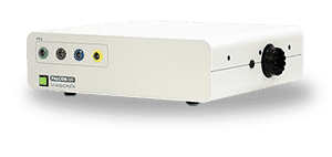
2 MHz Doppler Probe
High quality and ultra sensitive Doppler probe
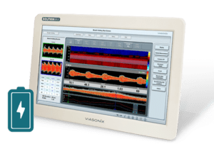
Using Dolphin for Stenosis Detection
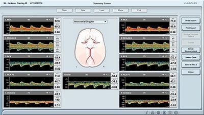
The Dolphin TCD system has a high-resolution spectral analysis that is key to analyze the waveform morphology in the various stenotic regions. The Doppler options include a simple and immediate toggle between different spectral color palettes for improved identification and visualization of Doppler systolic bruits.
A particularly unique and unprecedented ability of the Dolphin is to display the velocity profile across the arterial lumen when the ultrasound beam crosses the artery at an angle. The special Dolphin display option shows the continuous systolic and diastolic flows in the cross-section of the artery as a function of the cardiac cycle. Thus, under normal flow conditions, the velocity towards the walls is expected to be close to zero and maximal towards the middle of the lumen.
However, with a stenosis in the path of the flow, local turbulence may be visualized as retrograde flow in the proximity of the arterial walls and a sharp, high-velocity jet in the center.
The Dolphin TCD system also has exceptional multi-depth capabilities. The doppler waveforms from multiple different depths along the artery are measured and displayed simultaneously. Thus, with focal intracranial stenosis, the spectral Doppler in the proximal and distal regions of the stenosis, as well as at the focal stenosis location, can be analyzed both quantitatively and qualitatively.
Expected Results
The analysis of intracranial stenosis is both quantitative and qualitative by examining Doppler waveform morphology. It also requires measurements in the pre- and post-stenosis arterial segments.
There are several different criteria for assessing MCA stenosis. In general, the Mean velocity thresholds vary between > 80-120 cm/sec. According to one study, a focal Mean flow velocity > 80 cm/s, Peak velocity > 140 cm/s, and/or interhemispheric Mean flow difference > 30 cm/s, is suggestive of MCA stenosis.
The MCA stenosis severity is also estimated with TCD. The comparison of the velocities proximal and distal to the stenosis is a key factor.
When the post-stenotic to pre-stenotic velocity ratio is less than double, then the stenosis is considered about 30%. When the ratio increases to about 2, and the flow is disturbed with bruits, the stenosis severity is about 50%. A stenosis severity of 70% is expected when the ratio is 3-fold (or higher), and the flow is turbulent.
Selected Literature
Assessment: transcranial Doppler ultrasonography: Report of the Therapeutics and Technology Assessment Subcommittee of the American Academy of Neurology. Sloan MA et al., Therapeutics and Technology Assessment Subcommittee of the American Academy of Neurology. Neurology. 2004 May 11;62(9):1468-81
Accuracy of Transcranial Doppler Ultrasound Compared with Magnetic Resonance Angiography in the Diagnosis of Intracranial Artery Stenosis, Sandip Kumar Jaiswal et al., J Neurosci Rural Pract 2019;10:400–404
Hemodynamic consequences of cerebral vasospasm on perforating arteries: A phantom model study, Soustiel J, Levy E, Bibi R, Lukaschuk S, and Manor D. Stroke, 2001;32(3), p. 629-633
Transcranial Doppler Series Part IV: Case Studies, Heather A Nicoletto and Marilyn H. Burkman, Am J Electroneurodiagnostic Technol, 49:342-360, 2009
Transcranial Doppler: Technique and Common Findings (Part 1), Lokesh Bathala, Man Mohan Mehndiratta, and Sharma VK, Ann Indian Acad Neurol. 2013 Apr-Jun; 16(2): 174–179
Ultrasound Assessment of the Intracranial Arteries, Nabavi, Ritter, Otis, and Ringelstein, Introduction to Vascular Ultrasonography, By John Pellerito, MD and Joseph F Polak, 2012
MCA Stenosis, Robert A. Felberg, in Head Injury. Edited by Peter Reilly and Ross Bullock. Published in 1997 by Chapman & Hall, London
Neuro-ultrasonography, Ryan Hakimi, Andrei V. Alexandrov, and Zsolt Garami, Neurol Clin 38 (2020) 215–229
Disclaimer of Information & Content
The content of Viasonix Ltd. website is for information only, not advice or guarantee of outcome. Information is gathered and shared from reputable sources; however, Viasonix Ltd. Management is not responsible for errors or omissions in reporting or explanation. No individuals, including those under our active care, should use the information, resources or tools contained within this self-diagnosis or self-treat any health-related condition. Viasonix Ltd. Management gives no assurance or warranty regarding the accuracy, timeliness or applicability or the content.