


What is Photo-Plethysmography?
Photo-plethysmography (PPG) is a non-invasive testing method and is an essential tool in every physiological vascular testing examination.
The PPG sensors transmit infra-red waveforms to the skin and detect the signals that are reflected back to the sensor from the skin. The skin partially absorbs the transmitted signals, and the reflected signals are a function of this light absorption, which in turn is a function of the local blood perfusion. Therefore, the PPG waveforms reflect local and relatively shallow skin variations in blood flow.
The Photo-plethysmograph detects local blood volume changes and generates a pulsatile waveform, which is very similar to the Pulse Volume Recording (PVR) waveforms. However, PPG is based on optical technology, while PVR technology is based on a pressure-sensor measurement.
How to Perform PPG Measurement
Examiners usually use PPG measurements either to support the complete vascular diagnosis or as part of other physiological tests such as Toe Brachial Index (TBI) examinations, Venous Reflux test, TOS evaluation, Palmar Arch test, Raynaud’s examinations, and more.
Here is a detailed guide on how to perform the PPG test:
Step 1: Patient Preparation
- Position the patient comfortably in a relaxed state, sitting or preferably lying down.
- Explain the procedure to the patient to alleviate any concerns and ensure their cooperation.
Step 2: Sensor Placement
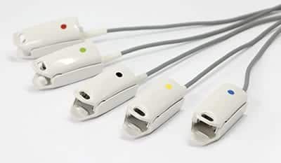
- Select the appropriate type of PPG sensor (Toe Clip, Finger Clip, or Disk sensor) according to the measurement you intend to perform.
- Apply the sensor to the target measurement site. For Toe Clips or Finger Clips, place them on the corresponding digits. Make sure to position the soft part of the finger or toe on top of the transparent windows of the PPG electronics. The transparent window is where the PPG sensors emit and detect the infrared waveforms, and it will pick up readings through the nails. For the Disk sensor, attach its electronics side to the skin with the dedicated adhesive sticker.
- Ensure proper attachment to avoid any movement artifacts during measurements.
Step 3: Equipment Setup
- Turn on the PPG measurement device.
- Configure the device settings as needed, including selecting the sensor type you’re using and adjusting the display settings (sweep time, amplitude display scale, etc.).
Step 4: Initiating a Measurement
- Start the PPG measurement on the device.
- The PPG sensors will emit infrared light that penetrates the skin, and the reflected signals from the blood perfusion will be detected.
Step 5: Freezing the Measurement
- The PPG waveforms are sensitive to movements. Pay attention to any abnormalities or changes in waveform amplitude, shape, or quality.
- Once 4 seconds of a stable waveform is observed, freeze the measurement.
Step 7: Interpreting Results
- Interpret the PPG measurements in the context of the patient’s medical history and other relevant diagnostic tests.
- Analyze the recorded PPG waveforms to identify any significant changes or patterns.
- Compare the PPG waveforms to established norms or previous measurements.
Step 8: Documentation and Reporting
- If the device provides an option, you can capture and save the recorded PPG waveforms for documentation and generate a report summarizing the measurement results, including waveform characteristics, abnormalities, and any relevant clinical observations.
This step-by-step guide for performing a PPG test is for informational purposes only. Healthcare professionals should rely on their expertise, clinical judgment, and institutional protocols for accurate administration and interpretation. Additional clinical information, patient history, physical examination, and other diagnostic tests may be necessary for comprehensive evaluation.
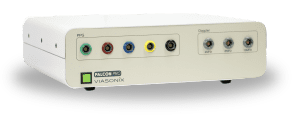
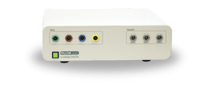
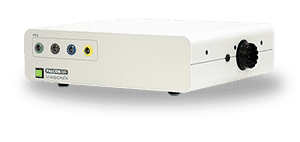
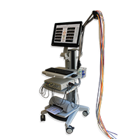
Using the Falcon for PPG Readings
The Falcon/Pro supports 5 independent and color-coded PPG ports, and the Falcon/Quad and Falcon/ABI+ models support 4 independent and color-coded PPG ports.
Here are some of the key benefits of utilizing the Falcon for PPG measurements:
1. High Precision and Accuracy: Advanced ultra sensitive PPG sensors to capture subtle changes in blood flow patterns and waveform characteristics.
2. Multiple Sensor Configurations: Diverse range of PPG sensor configurations, including Toe Clip, Finger Clip, and Disk sensors for measurements in various anatomical locations. The Disk PPG sensor can be attached to the skin with a special dedicated PPG adhesive sticker, which is practical for measurements on the calf or the tip of the toes.
3. Comprehensive Physiological Testing: Various physiological testing protocols, making it a versatile tool for vascular assessment such as Toe Brachial Index (TBI) measurements, Venous Reflux tests, Thoracic Outlet Syndrome (TOS) evaluations, and Palmar Arch assessments.
4. Simultaneous Multi-Site Measurements: Multiple independent and color-coded PPG ports to perform simultaneous measurements on different fingers or toes. This is especially useful in scenarios like quick Raynaud’s assessments or TBI studies, enhancing both efficiency and patient comfort.
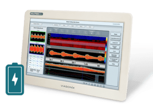
5. Customizable Configuration: The Falcon photoplethysmography system provides complete control over waveform display settings, including sweep time, amplitude display scale, and signal filtering.
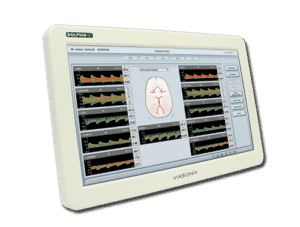
6. Enhanced Diagnostics with Summary Screens: Dedicated summary screens enriched with schematic pictures facilitate clear and comprehensive test assessments.
7. Real-time Data Presentation: Display of PPG waveforms in real time and offline replay of the entire PPG recording.
Expected Results
PPG measurements are widely used in various physiological test protocols, and in most cases, the main objective is to qualitatively diagnose changes in waveform quality or amplitude under specific conditions. For example, a sharp decrease in waveform amplitude during a TOS test when testing a particular position, a flattening of the PPG curve during vessel compression as part of the Palmar Arch test, or a very long recovery time after ice water immersion during a Raynaud’s Syndrome examination.
In addition, PPG waveforms help to determine systolic blood pressure during TBI or ABI diagnosis. A proximal pressure cuff is inflated until the distal PPG waveform flattens with total occlusion. Then, during the cuff deflation process, the pressure at which the PPG waveform reappears for the first time is considered as the systolic blood pressure at the measurement site.
Selected Literature
Overview of Peripheral Arterial Disease of the Lower Extremity, Ali F. AbuRahma and John E. Campbell, Noninvasive Vascular Diagnosis, A.F. AbuRahma (ed.), Springer International Publishing AG 2017, Ch 21, pp 291-318
Comparison of two methods based on photoplethysmography for the diagnosis of peripheral arterial disease, Christian Høyer et al., Scandinavian Journal of Clinical and Laboratory Investigation, 2017
The effect of percutaneous transluminal angioplasty of superficial femoral
artery on pulse wave features, Mikko Peltokangas et al., Computers in Biology and Medicine 96 (2018) 274–282
Nonimaging Physiologic Tests for Assessment of Lower Extremity Arterial Disease, Marsha M. Neumyer, in “Introduction to Vascular Ultrasonography”, Ed. Pellerito and Polak, Elsevier Health Sciences, 2012, Ch 14, pp 244-261
Peripheral vascular disease assessment in the lower limb: a review of current and emerging non‑invasive diagnostic methods; Shabani Varaki et al, BioMed Eng OnLine (2018) 17:61
Photoplethysmographic Venous Refilling Times Following Ultrasound Guided Foam Sclerotherapy for Symptomatic Superficial Venous Reflux: Relationship with Clinical Outcomes, Darvall et al., Eur J Vasc Endovasc Surg (2010) 40, 267e272
Disclaimer of Information & Content
The content of Viasonix Ltd. website is for information only, not advice or guarantee of outcome. Information is gathered and shared from reputable sources; however, Viasonix Ltd. Management is not responsible for errors or omissions in reporting or explanation. No individuals, including those under our active care, should use the information, resources or tools contained within this self-diagnosis or self-treat any health-related condition. Viasonix Ltd. Management gives no assurance or warranty regarding the accuracy, timeliness or applicability or the content.